
Computational Surgical Oncology (CSO) group
German version
The Computational Surgical Oncology (CSO) group conducts translational research in artificial intelligence and data science.
Core competence is computer vision, using high-resolution images as input for neural networks. We use deep learning with convolutional neural networks (CNNs).
We do projects in oncological surgery, oncology, and surgery. This approach advances personalized medicine, especially personalized cancer therapy.

Our Vision
To personalize therapy decisions by turning all digital patient data into biomarkers with the help of artificial intelligence.
Often there are multiple equivalent therapies for a patient. Personalized cancer therapy predicts which patients are more likely to respond to one of these specific therapies. Biomarkers, especially in cancer medicine, are used for this prediction. To date, few biomarkers are measured to individualize therapy decisions, and optional targeted therapy is usually initiated based on these biomarkers.
One example of such a biomarker targeting oesophageal cancer is the human epidermal growth factor receptor 2 (HER2). HER2, a member of the EGFR family, is localized on the cell membrane and accelerates tumor growth. As a biomarker, the HER2 status determines the potential administration of antibody therapy with trastuzumab. This therapy is recommended for palliative esophageal carcinoma if HER-2 expression is present, i.e., a positive HER-2 status.
The more biomarkers used, the more treatment decisions can be individualized. The idea of the Computational Surgical Oncology group is to digitize all available patient data and use them as biomarkers for individual therapy decisions. With the help of neural networks, the resulting complex and extensive data can be mastered and transformed into personal therapy decisions.
Of particular interest are pathological image data, such as morphological H&E staining, which contain more information than is visible to the bare eye. Thus, it has been possible to predict molecular mutations based on H&E data.
The Computational Surgical Oncology group uses digital H&E staining (about 10 Gb per image) to predict the response to cancer therapies. In the future, this will allow individualized decisions as to whether a patient should have upfront surgery or neoadjuvant therapy.
The type and number of biomarkers will be gradually increased for the personalization of cancer therapy. Clinical data will be combined with H&E stainings to optimize the prediction of response to therapy. Later, radiological imaging such as computed tomography (CT) will also be integrated as biomarkers. In addition to choosing the optimal individual therapy, artificial intelligence can be used to determine the prognosis for cancer.
Digital biomarkers support important surgical decisions during operations. In addition to cancer therapy, the focus of the Computational Surgical Oncology group is on AI-supported optimization of surgical strategy. Biomarkers can, for example, help in planning surgery to select the most suitable surgical procedure for an individual patient. Artificial intelligence methods also assess the surgical risk on an individual basis.
Current projects
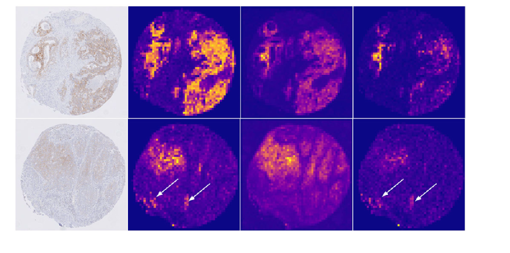
Predicting the HER2 status in esophageal cancer from tissue microarrays using convolutional neural networks
We aim to develop a deep-learning method to screen microscopic images of esophageal cancer for the presence of HER2 overexpression. Our solution enables direct automated detection of HER2 in immunohistochemically stained tissue sections without the need for manual assessment and additional costly in situ hybridization (ISH) assays.
We trained neural networks (CNNs) on a large dataset of 1,602 patient samples and tested them on an independent set of 307 patient samples. We incorporated an attention mechanism into the CNN architecture to identify the tissue regions that the network identifies as important for the prediction.
Our method has an accuracy of 0.94, a precision of 0.97, and a recognition of 0.95. Importantly, our approach provides accurate predictions in cases that pathologists can not resolve without additional FISH testing.
Team
Dr. Jin-On Jung
Dr. Karl Knipper
Dr. Leandra Lukomski